Home » Without Label » Muscle Chart Back / Male Back Muscles Chart - Human Anatomy Body - Human ... / Creatine research more than a sports supplement read more….
Muscle Chart Back / Male Back Muscles Chart - Human Anatomy Body - Human ... / Creatine research more than a sports supplement read more….
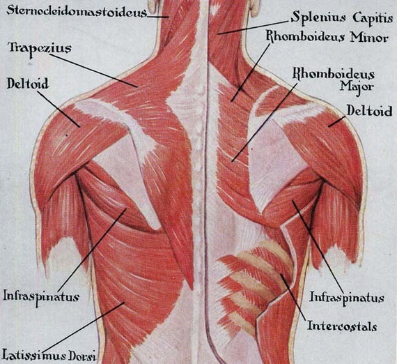
Muscle Chart Back / Male Back Muscles Chart - Human Anatomy Body - Human ... / Creatine research more than a sports supplement read more….. Muscle spasms (contraction or stiffening of the back muscles) muscles that feel tight; The most common type of back pain is muscle pain—also called muscle strain or soft tissue strain. The deltoid, teres major, teres minor, infraspinatus, supraspinatus (not shown) and subscapularis muscles (not shown) all extend from the scapula to the humerus and act on the shoulder joint. A large muscle group in the shoulder, neck and upper back that pulls the head and shoulders backward. The teres major muscle originates on the outer (lateral) edge of the scapula and attaches to the humerus.
Creatine is now proving to be one of the most potent muscle growth accelerators giving excellent muscle mass increase and phenomenal strength increases order yours today. There are three different muscle groups found in the back: Body muscle anatomy human body anatomy human anatomy and physiology anatomy study anatomy reference human skeleton anatomy vie simple muscle structure medical anatomy. A strain can be an injury to a tendon attachment from muscle to bone. Out of these, the cookies that are categorized as necessary are stored on your browser as they are essential for the working of basic functionalities of the website.
Muscles of the Shoulder and Back Laminated Anatomy Chart from cdn11.bigcommerce.com Creatine is now proving to be one of the most potent muscle growth accelerators giving excellent muscle mass increase and phenomenal strength increases order yours today. The superficial group, the deep group, and the intermediate group. This website uses cookies to improve your experience while you navigate through the website. Muscle anatomy of forearm 12 photos of the muscle anatomy of forearm anatomy of forearm. Latissimus dorsi a back muscle that pulls the arm down and back. Loss of control of the bowel or bladder and retention of urine may. The back consists of the spine, spinal cord, muscles, ligaments, and nerves. Nerves in your lower back.
A large muscle group in the shoulder, neck and upper back that pulls the head and shoulders backward.
The back consists of the spine, spinal cord, muscles, ligaments, and nerves. The two trapezius muscles extend from the backbone and base of the skull, across the back and shoulders to join the scapula and the clavicle. We've created a free trigger point chart, which includes fybromyalgia treatment and reflexology information. This website uses cookies to improve your experience while you navigate through the website. The back's muscles start at the top of the back (named the cervical vertebrae) and go to the tailbone (also named the coccyx). This diagram depicts back shoulder muscles.human anatomy diagrams show internal organs, cells, systems, conditions, symptoms and sickness information and/or tips for healthy living. Muscle spasms (contraction or stiffening of the back muscles) muscles that feel tight; The deep muscles of the back fit into or affix parts of themselves to the grooves in the spinous. Some of the links in the post above are affiliate links.. Anatomy of the upper back. The trapezius and latissimus dorsi muscles connect the upper limb to the vertebral column. These structures work together to support the body, enable a range of movements, and send messages from the. To download your free copy click the link.
The trapezius and latissimus dorsi muscles connect the upper limb to the vertebral column. There are a few warning signs, however, that may indicate serious spinal problems. For more anatomy content please follow us and visit our website: Body muscle anatomy human body anatomy human anatomy and physiology anatomy study anatomy reference human skeleton anatomy vie simple muscle structure medical anatomy. Loss of control of the bowel or bladder and retention of urine may.
Muscles Back Posterior Human Anatomy Vintage Medical Chart from img.etsystatic.com Most of the time, back muscle pain is diagnosed then treated with little more than a prescription of rest, painkillers and muscle relaxants. The back consists of the spine, spinal cord, muscles, ligaments, and nerves. Five pairs of lumbar spinal nerves labeled l1 to l5 branch off your spinal cord and exit through small holes between the vertebrae. Creatine research more than a sports supplement read more…. Muscles connect to the vertebrae and bones via ligaments, flexible bands of fibrous tissue. The pelvic floor muscles also help increase this pressure, which provides stability to the spine and trunk. Muscle charts of the human body for your reference value these charts show the major superficial and deep muscles of the human body. Artery) p.134 accessory nerve p.
This diagram depicts back shoulder muscles.human anatomy diagrams show internal organs, cells, systems, conditions, symptoms and sickness information and/or tips for healthy living.
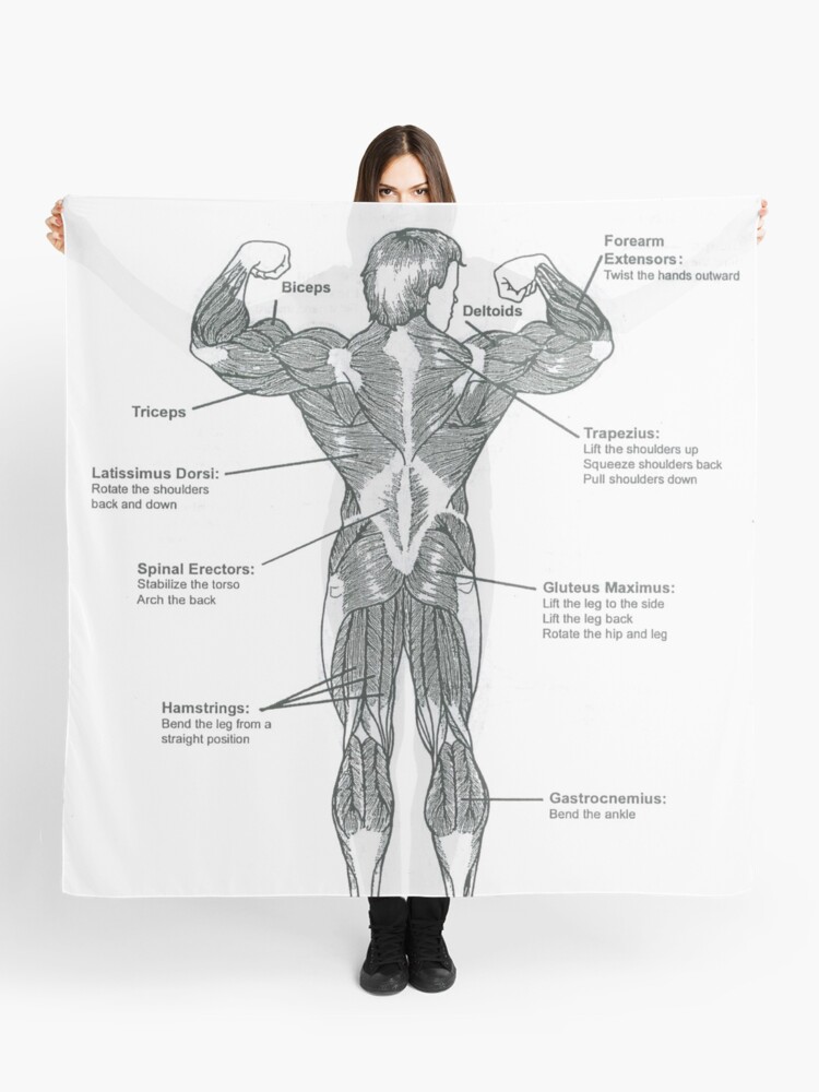
They lift and tilt head and lift or steady the shoulders. A strain can be an injury to a tendon attachment from muscle to bone. Muscles of back (trapezius, latissimus dorsi) There are three different muscle groups found in the back: For more anatomy content please follow us and visit our website: The deep muscles of the back fit into or affix parts of themselves to the grooves in the spinous. Loss of control of the bowel or bladder and retention of urine may. Some of these muscles are quite large and cover broad areas. The part of the nerve that emerges out of the spine is called the nerve root. Others, like sumo deadlifts, have been shown in emg studies—and in the trenches—to focus more on other muscle groups than the back. Anatomy of the upper back. Your clients will thank you for it! Claim your free copy of the client back care guide today.
The deltoid, teres major, teres minor, infraspinatus, supraspinatus (not shown) and subscapularis muscles (not shown) all extend from the scapula to the humerus and act on the shoulder joint. Both the deltoid and the trapezius are firmly attached to the spine of the scapula. Related posts of lower back muscle chart muscle anatomy of forearm. The deep muscles of the back fit into or affix parts of themselves to the grooves in the spinous. Five pairs of lumbar spinal nerves labeled l1 to l5 branch off your spinal cord and exit through small holes between the vertebrae.
Muscle Chart Back : Muscles German Names Chart Muscular ... from ih1.redbubble.net 1) make midline incision along spines of vertebrae 2) extend from There are a few warning signs, however, that may indicate serious spinal problems. Both the deltoid and the trapezius are firmly attached to the spine of the scapula. Nerves in your lower back. The teres majo r muscles work with the rotator cuff muscles to stabilize. Latissimus dorsi a back muscle that pulls the arm down and back. The quick answer to this question is the muscles of the lower back are the multifidus, longissimus, spinalis, and quadratus lumborum. Claim your free copy of the client back care guide today.
Muscle spasms (contraction or stiffening of the back muscles) muscles that feel tight;
The deep muscles of the back fit into or affix parts of themselves to the grooves in the spinous. Latissimus dorsi a back muscle that pulls the arm down and back. For more anatomy content please follow us and visit our website: Muscle anatomy of forearm 12 photos of the muscle anatomy of forearm anatomy of forearm. Three types of back muscles that help the spine function are extensors, flexors and obliques. There are a few warning signs, however, that may indicate serious spinal problems. We've created a free trigger point chart, which includes fybromyalgia treatment and reflexology information. These muscles include the large paired muscles in the lower back, called erector spinae, which help hold up the spine, and gluteal muscles. The vast majority of back problems improve on their own or with nonsurgical treatment. The back consists of the spine, spinal cord, muscles, ligaments, and nerves. Some of the links in the post above are affiliate links.. Muscles connect to the vertebrae and bones via ligaments, flexible bands of fibrous tissue. The trapezius and latissimus dorsi muscles connect the upper limb to the vertebral column.